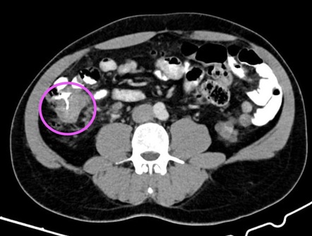
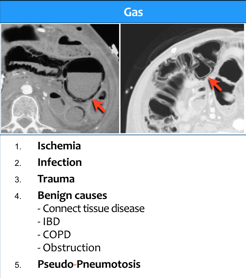
2124 Complications of typhlitis on CT include pneumatosis pneumoperitoneum and pericolic fluid. The borders are shouldering unlike in diverticulitis where the borders are tapering figure.
Ct Scan Bowel Wall Thickening Ct Scan Machine
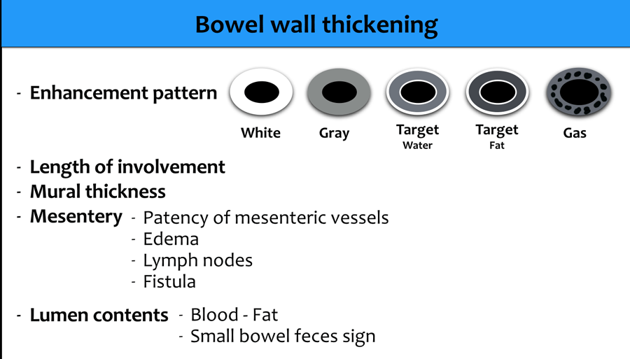
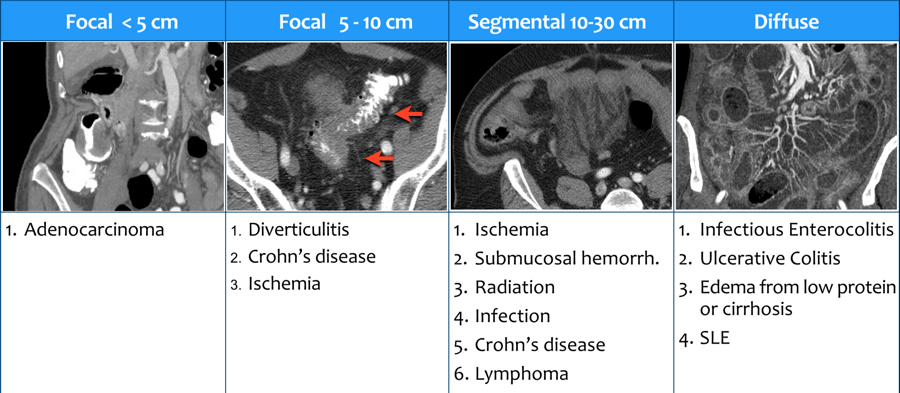
Mural thickening may be diffuse segmental or focal.

Mural thickening meaning. Thickening of the gallbladder wall is a relatively frequent finding at diagnostic imaging studies. Focal thickening may be caused by tumours or by inflammatory conditions and distinguishing between the two conditions should be attempted. The process of becoming thicker or of making something become thicker.
It may occur in any segment of the esophagus although it is more common distally. CT especially after the advent of multidetector helical scanning has led to the increased detection of subtle gastrointestinal tract abnormal findings. AimsBackground Further endoscopic evaluation often is suggested when BWT is identified on CT imaging Is there clinical correlation.
Esophagitis is more likely than esophageal carcinoma. Thickening of the bowel wall is considered focal when it extends less than 5 cm 3 11. Patency of the mesenteric vessels.
However edema congestive heart failure cirrhosis of the liver infections parasitic infestations and inflammation can also cause intestinal thickening. Cat scan of belly revealed colon wall thickening and i have been having awful belly pain. In addition to the clinical presentation analysis of the wall symmetry degree of thickening and perienteric abnormalities provides additional information for the.
Thickening of the bowel wall may be focal 40 cm in extension. The wall thickening can be isodense or hypoattenuating which is indicative of edema hemorrhage or necrosis. Regular symmetric and homogeneous wall thickening is more.
Blockages in the carotid artery of a certain severity may increase ones risk of stroke. When its time to urinate the bladder wall muscles tighten to help push urine out through the urethra. Mural thickening may be diffuse segmental or focal.
Degree of mural thickening. For those carotid arteries without a blockage some physicians measure the intima the inner-most lining of the artery and derive a value called the imt or intimal-medial thickness. Mural thickening of intestine causing obstruction in Chinese.
All these features will be discussed in detail in the following paragraphs. When the bladder is filling with urine the muscles in the bladder wall relax. Intravenous contrast material administration is helpful in.
What does that mean. Universal Images Group EditorialUniversal Images GroupGetty Images. 1 Bowel wall thickening is one of the important gastrointestinal tract abnormalities increasingly being detected on CT.
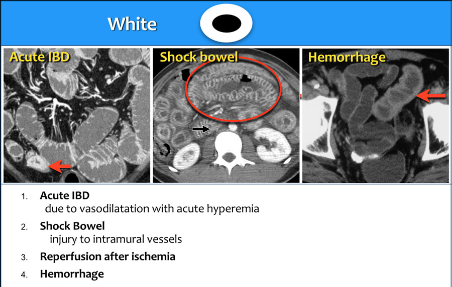
There are many pathophysiologic events that can cause a white attenuation The CT findings are. Inflamation Answered by Dr. Lenght of bowel wall involvement 5 cm involvement Adenocarcinoma usually presents as a short segment of bowel wall thickening.
The Doctor needs to review all the information CAT scan. 1 The bowel wall thickening could be a manifestation of underlying inflammatory infectious ischemic or neoplastic. 22 The cecum may also demonstrate distension or dilatation with hyperattenuating adjacent fat and thickening of fascial planes indicating pericolonic and mesenteric inflammation.
MD Consult states that the most common reason people experience thickening of the colon wall is diverticulitis. Circumferential and diffuse mural thickening with submucosal edema indicating pericolonic and mesenteric inflammation andor fat stranding are the common CT findings in patients with metastases to small bowel segments Figure 28-4 or neoplastic conditions and the presence of.
Introduction Bowel wall thickening BWT is an increasing recognised entity seen on CT. Intravenous contrast material administration is helpful in the CT evaluation of esophageal mural thickening. Mural thickening is secondary to edema and appears as a sonolucent line between two echogenic lines in the gallbladder wall.
Click for more detailed Chinese translation meaning. Is gallbladder wall thickening dangerous. Advances in technology and accumulated experience in image interpretation even the most subtle changes affecting the bowel are now being detected.
Perienteric fat stranding disproportionally more severe than the degree of wall thickening suggests an inflammatory condition. It may occur in any segment of the esophagus although it is more common distally. Imaging description Esophageal mural thickening is a nonspecific finding by CT chest.
Focal irregular and asymmetrical thickening of the bowel wall suggests a malignancy.

The Radiology Assistant Gallbladder Wall Thickening

Significance Of Ileal And Or Cecal Wall Thickening On Abdominal Computed Tomography In A Tropical Country Kumar 2019 Jgh Open Wiley Online Library
The Radiology Assistant Ct Pattern Of Bowel Wall Thickening
Https Www Ajronline Org Doi Pdf 10 2214 Ajr 176 5 1761105
The Radiology Assistant Ct Pattern Of Bowel Wall Thickening
Bowel Wall Thickening Radiology Reference Article Radiopaedia Org

Five Cte Parameters A Mural Hyperenhancement Defined As Segmental Download Scientific Diagram
The Radiology Assistant Ct Pattern Of Bowel Wall Thickening

Bowel Wall Thickening Radiology Reference Article Radiopaedia Org

Ileocecal Thickening Clinical Approach To A Common Problem Agarwala 2019 Jgh Open Wiley Online Library

The Radiology Assistant Gallbladder Wall Thickening

Bowel Wall Thickening On Kub Youtube

Ct Scan Of The Lower Abdomen Demonstrates Concentric Mural Wall Download Scientific Diagram

Bowel Wall Thickening Radiology Reference Article Radiopaedia Org
Pdf Bowel Wall Thickening At Ct Simplifying The Diagnosis

Ileocecal Thickening Clinical Approach To A Common Problem Agarwala 2019 Jgh Open Wiley Online Library
The Radiology Assistant Ct Pattern Of Bowel Wall Thickening

Pdf Bowel Wall Thickening At Ct Simplifying The Diagnosis
Pdf Bowel Wall Thickening At Ct Simplifying The Diagnosis

